Assignment:
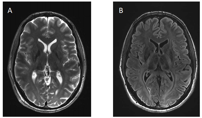
Q1. Examine the images A and B acquired at 7T below. For each image, discuss the origin of the contrast observed in the CSF, white matter (WM) and grey matter (GM). As part of your answer:
a. state the type of weightings in each image and give brief reasoning
b. suggest a sequence that could produce this weighting for each image
c. suggest possible range of TE, TR, and TI (if appropriate) values that might produce these images given the following properties:
• average T1 at 7T: CSF 4sec, WM 1.4sec and GM 2sec
• approx. T2: CSF 500ms, WM 40ms and GM 50ms

Q2. Image B above was acquired with the following acquisition parameters: FOV 23cm x 23cm, receiver BW 78,080Hz, pixel size 0.72mm x 0.72mm.
a. Draw a representative sequence diagram for this pulse sequence.
(Do not copy and paste a graphic from a text or the web - draw by freehand if necessary and insert a scan or photograph.)
Include:
• All necessary RF pulses
• All gradient pulses
• Echo signals
• Indicate TR and TE and other critical parameters if appropriate
• Label each line of the sequence
b. Calculate the dwell time for the acquisition
c. Calculate the read gradient strength used in mT/m
d. Calculate the image matrix size